Optical Coherence Tomography (OCT) of the Optic Nerve
Fundamental for expert and accurate monitoring and early diagnosis of glaucoma.
What does it entail?
OCT (Optical Coherence Tomography) in glaucoma is a non-invasive test of the back of the eye, which provides high-resolution scans of the retinal layers.
For which diseases is it used?
OCT of the optic nerve is used to diagnose and monitor various neuropathies, including: glaucoma, optic neuritis, optic disc drusen, etc.
Glaucoma is a disease of the optic nerve, consisting of a gradual and characteristic loss of the nerve fibres that make up the optic nerve. There is a high variability in the nerve fibre count in the optic nerves of different people, varying from 700,000 to 1,700,000 (Jonas JB, Schmidt AM, et al. Human optic nerve fiber count and optic disc size. IOVS), but when glaucomatous damage begins, the fibres become atrophied in a specific pattern in all people and this is exactly what OCT is looking for.
What OCT equipment does ICO have?
Here at ICO, we have the Cirrus spectral-domain OCT (manufactured by Carl Zeiss), which has higher resolution and scan speed than the earlier Stratus time-domain OCT. This technology allows the SD-OCT to obtain retinal images some 50 times faster than the TD-OCT (18,000- 27,000 axial scans per second).
What are the technical principles of OCT?
OCT is based on the optical principle of interferometry. It uses an infrared light source that enters the eye, reflects on the retina and produces an interference when scattered, "translated" by the OCT into a retinal thickness map. There is a colour code: cold colours (blue, black) represent thinner tissue, while warm colours (red, yellow, white) represent thicker tissue.
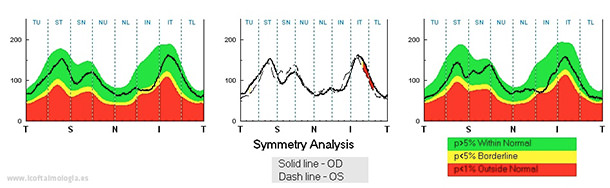
OCT in glaucoma measures the retinal region that is very close to the optic nerve (layer of peripapillary nerve fibres) and compares it to a database of people of the same age who do not have glaucoma.
How can it be useful?
Your ophthalmologist can interpret these results and explain them to you. Sometimes, in a very small percentage of cases, there are false positives (i.e. the software's ¨automated¨ analysis indicates that there is damage to the nerve but this damage does not really exist) and false negatives (the ¨automated¨ analysis indicates normality but there is evidence of glaucomatous damage in the visual field test or on the black-and-white photo of the retina). This is why several tests are required to diagnose and monitor glaucoma, such as visual field, red-free retinography, OCT, intraocular pressure and direct examination of the nerve by the specialist.
One very useful application of OCT in glaucoma is the ability to assess disease progression by performing periodic studies with the pre-installed inter-scan change analysis software.
OCT is a very useful tool for discovering very small optic nerve defects and for diagnosing glaucoma at a very early stage, before visual field impairment is evident (which we call pre-perimetric glaucoma).
In the days before optic nerve measurement technology, the definition of glaucoma was based on specific visual field impairment. Today, however, the disease can be diagnosed much sooner and treatment can be started earlier.